Understanding Knee Ligament Anatomy: Your Complete Guide to Knee Stability
Your knee is one of the most complex joints in your body, and I see patients every day who want to understand exactly what's happening inside this remarkable structure. As an orthopedic surgeon in Franklin, Tennessee, I've treated thousands of knee injuries over the years, and I can tell you that understanding your knee ligament anatomy is the first step toward better knee health.
Think of your knee ligaments as the strong cables that keep your knee stable and moving properly. When patients ask me "What exactly are ligaments doing in my knee?" I explain that they're like the guy-wires that hold up a radio tower - without them, everything would collapse. Your knee has several key ligaments working together 24/7 to keep you walking, running, and living your active life.

What Are Knee Ligaments and Why Do They Matter?
Knee ligaments are tough, fibrous bands of tissue that connect your thigh bone (femur) to your shin bones (tibia and fibula). I tell my patients to imagine them as nature's duct tape - they hold bones together while still allowing controlled movement.
Your knee actually houses multiple ligament systems, each with a specific job:
The Four Major Players:
- Anterior Cruciate Ligament (ACL)
- Posterior Cruciate Ligament (PCL)
- Medial Collateral Ligament (MCL)
- Lateral Collateral Ligament (LCL)
The Supporting Cast:
- Patellar ligament (technically a tendon, but everyone calls it a ligament)
- Anterolateral ligament (ALL) - a newer discovery
- Transverse ligament
- Various meniscal attachments
After examining thousands of knees, I can tell you that each ligament has evolved to handle specific stresses. When one gets injured, the others often have to work overtime, which is why knee injuries can be so complex.
The ACL: Your Knee's Most Famous Ligament
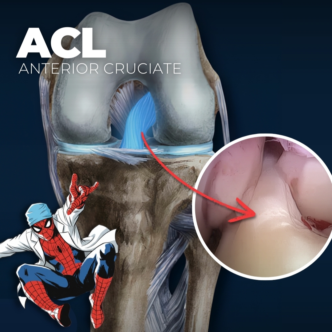
The ACL gets all the headlines, and for good reason. This ligament controls your knee's rotational stability and prevents your shin bone from sliding too far forward. I see about 200,000 ACL injuries nationwide every year, making it the superstar of sports medicine.
What Makes the ACL Special
The ACL sits right in the center of your knee, crossing over the PCL to form an "X" shape. It's about 32-34 millimeters long and handles some of the most complex movements your knee makes. Every time you pivot, cut, or change direction quickly, your ACL is working hard.
When ACL Injuries Happen
In my practice, I see ACL tears most often in athletes who play sports involving sudden direction changes - basketball, soccer, football, and skiing. Women are actually 4-6 times more likely to tear their ACL than men, due to differences in anatomy, hormones, and muscle activation patterns.
The classic ACL injury happens when you:
- Land awkwardly from a jump
- Stop suddenly while running
- Change direction rapidly with your foot planted
- Take a direct blow to the knee
Most patients describe hearing a "pop" followed by immediate swelling and the feeling that their knee won't support them properly.
The PCL: Your Knee's Unsung Hero
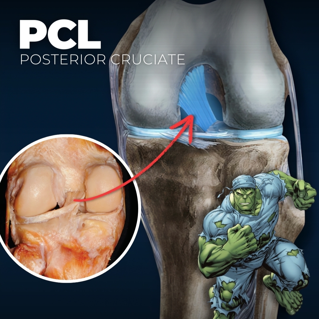
Here's something that might surprise you - your PCL is actually stronger than your ACL. Much stronger. I call it the "Hulk" of knee ligaments because it's nearly twice as thick and handles enormous forces without complaining.
The PCL's Tough Job
The PCL prevents your shin bone from sliding backward and provides stability when your knee is bent. Unlike the ACL, which tears relatively easily, PCL injuries are much less common because this ligament is built like a steel cable.
PCL Injury Signs
PCL injuries usually require significant trauma - car accidents, direct blows to the front of the knee, or severe hyperextension. Patients often don't realize they've injured their PCL because the knee might still feel relatively stable, unlike ACL injuries.
The Collateral Ligaments: Your Knee's Side Guards
The MCL and LCL work like bookends, preventing your knee from bending sideways in ways it shouldn't. I see these injuries frequently in contact sports and skiing accidents.
MCL: The Flat Protector
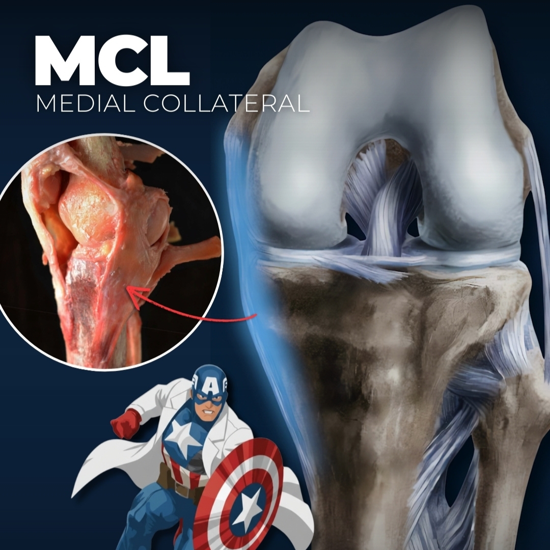
Your MCL is wide and flat, like a shield protecting the inside of your knee. It's actually made up of six distinct bands that work together. Here's some good news - the MCL heals better than most other knee ligaments because it has excellent blood supply.
MCL injuries typically happen when:
- Your knee gets hit from the outside (like a football tackle)
- You land with your knee bent inward
- You experience a twisting motion with your foot planted
LCL: The Precision Specialist
The LCL is much thinner than the MCL - more like a strong cord than a flat band. It protects the outside of your knee and is the only major knee ligament that doesn't attach to the meniscus. This independence actually makes it less likely to be injured in combination with other structures.

Lesser-Known but Important Ligaments
The Patellar Ligament: An Identity Crisis
Here's a fun fact that medical students always find confusing - the "patellar ligament" isn't technically a ligament at all. It's actually part of your quadriceps tendon that connects your kneecap to your shin bone. But since it connects bone to bone, everyone calls it a ligament.
This structure is incredibly important for knee extension. Every time you straighten your leg or kick something, you're using your patellar ligament to transfer power from your quadriceps muscle to your lower leg.
The Anterolateral Ligament: The New Kid on the Block
The ALL wasn't even officially described until 2013, which shows you how much we're still learning about knee anatomy. This tiny ligament (only 1-2.5 millimeters thick) helps with rotational control, and not everyone even has one.
What makes the ALL interesting is its variability - it can be anywhere from 30 to 59 millimeters long, and its presence varies between people. Research suggests it might play a role in some ACL reconstruction outcomes.

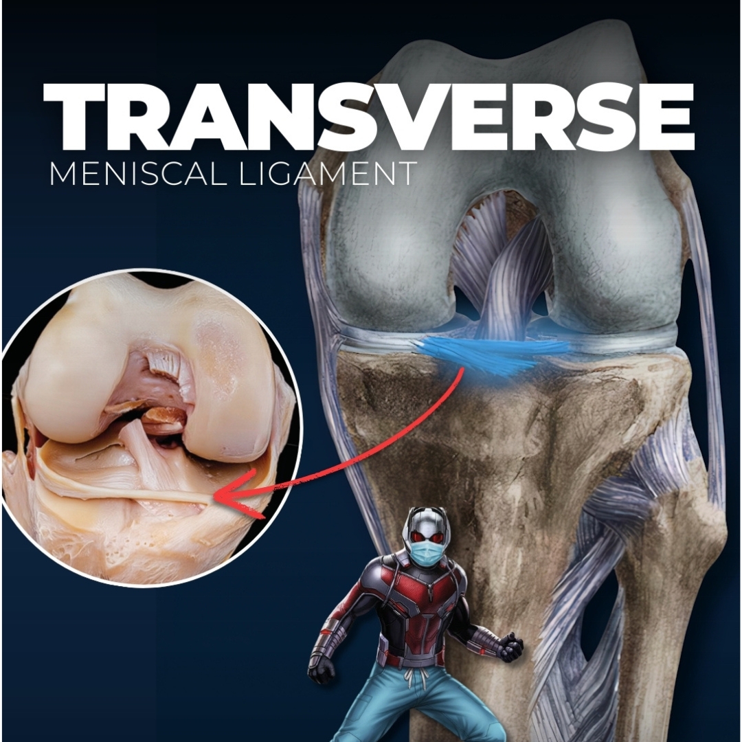
The Transverse Ligament: Small but Mighty
At only 20 millimeters long, the transverse ligament connects your menisci together. I think of it as the ligament world's version of Ant-Man - tiny but surprisingly important for knee stability.
When Should You Worry About Knee Ligament Injuries?
After 15 years of treating knee problems, I can tell you the warning signs that mean you need professional evaluation:
Seek immediate care if you experience:
- A "pop" in your knee followed by severe pain
- Knee swelling that develops within hours
- Your knee feels unstable or gives way
- You can't bear weight comfortably
- Your knee locks or gets stuck in one position
Schedule an appointment soon if you have:
- Persistent knee pain lasting more than a few days
- Swelling that doesn't improve with rest and ice
- A feeling that your knee might give out
- Difficulty with stairs or pivoting movements
Don't try to "tough it out" with knee injuries. Early treatment almost always leads to better outcomes.
Treatment Options for Knee Ligament Injuries
The good news is that we have excellent treatment options for most knee ligament injuries. Treatment depends on which ligament is injured, how severe the injury is, and your activity level.
Non-Surgical Treatment
Many ligament injuries can heal with conservative treatment:
- Rest and activity modification
- Physical therapy to strengthen surrounding muscles
- Bracing for additional support
- Anti-inflammatory medications
- Sometimes injections for pain and inflammation
Surgical Options
Some injuries require surgical repair or reconstruction:
- ACL reconstruction using tendon grafts
- PCL reconstruction for severe injuries
- MCL repair or reconstruction
- Multi-ligament reconstruction for complex injuries
I always tell patients that surgery isn't about getting back to where you were - it's about getting back to where you want to be. We tailor every treatment plan to your specific goals and lifestyle.
Protecting Your Knee Ligaments
Prevention is always better than treatment. Here's what I recommend to my patients in Franklin who want to keep their knees healthy:
Strengthen the Right Muscles:Your quadriceps, hamstrings, and glutes all work together to protect your knee ligaments. Weak muscles mean your ligaments have to work harder.
Improve Your Movement Patterns:Poor jumping and landing mechanics put excessive stress on ligaments. Sports-specific training can teach you safer movement patterns.
Don't Ignore Pain:That nagging knee pain isn't something to push through. Early intervention prevents small problems from becoming big ones.
Stay Flexible:Tight muscles can alter knee mechanics and increase ligament stress. Regular stretching keeps everything moving properly.
Recovery and Rehabilitation: What to Expect
Ligament healing takes time, and I always prepare patients for a process, not a quick fix. Here's what you can generally expect:
MCL injuries: Often heal well with 6-8 weeks of conservative treatmentACL injuries: Typically require surgery followed by 6-9 months of rehabilitationPCL injuries: May heal without surgery but often need 3-6 months of therapyCombined injuries: Can take 9-12 months for full recovery
The key to successful recovery is patience and compliance with your rehabilitation program. I've seen too many patients rush back too quickly and end up with recurring problems.
Living with Knee Ligament Injuries
Many patients ask me, "Will I ever be normal again?" The honest answer is that most people can return to their previous activity level, but it requires commitment to the treatment process.
Some patients do need to modify their activities. High-level pivoting sports might not be realistic after certain injuries, but that doesn't mean you can't stay active. I work with patients to find activities they can do safely and enjoyably.
The Bottom Line on Knee Ligament Anatomy
Your knee ligaments are remarkable structures that work together to provide stability while allowing complex movement. Understanding how they work helps you recognize when something's wrong and why certain treatments are necessary.
Remember, knee ligament injuries are often preventable with proper conditioning and technique. When injuries do occur, early treatment and proper rehabilitation give you the best chance for a full recovery.
If you're experiencing knee pain or instability in the Franklin area, don't wait to seek evaluation. As an orthopedic surgeon who specializes in knee problems, I'm here to help you understand your options and get back to the activities you love.
Ready to take the next step? Contact Dr. Cory Calendine's office in Franklin, Tennessee, to schedule a consultation. We'll evaluate your knee thoroughly and create a treatment plan that fits your lifestyle and goals.
This information is for educational purposes only and should not replace professional medical advice. Always consult with a qualified orthopedic surgeon for proper diagnosis and treatment of knee injuries.


.png)
