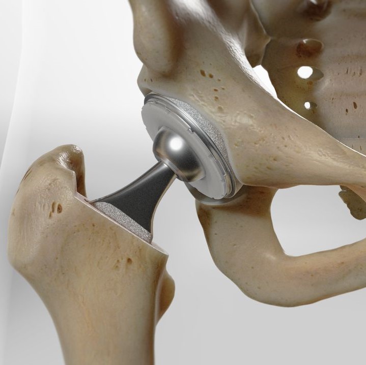
How is Direct Anterior Approach Hip Replacement Done? A Complete Surgical Guide
Hip replacement surgery has evolved dramatically over the past decades, with the Direct Anterior Approach (DAA) emerging as one of the most innovative and patient-friendly techniques available today. This minimally invasive procedure offers faster recovery times, reduced pain, and improved outcomes compared to traditional hip replacement methods.
If you're considering hip replacement surgery or simply want to understand this revolutionary technique, this comprehensive guide will walk you through exactly how Direct Anterior Approach hip replacement is performed in the operating room.
What is Direct Anterior Approach Hip Replacement?
The Direct Anterior Approach is a minimally invasive surgical technique for total hip replacement that accesses the hip joint from the front of the body. Originally described by Carl Heuter in 1881 and later popularized by Smith-Peterson, this approach has gained significant popularity in recent years due to its muscle-sparing benefits.
Unlike traditional hip replacement methods that require cutting through major muscle groups, DAA utilizes the natural space between muscles to reach the hip joint, preserving muscle integrity and accelerating recovery.
Pre-Surgical Preparation and Patient Positioning
Operating Room Setup
The surgical process begins with careful patient positioning, which is crucial for successful DAA hip replacement. The patient is positioned supine (lying on their back) on either a specialized fracture table or a standard operating room table.
When using a regular operating table, the patient's hip is strategically placed over the table break, which can be flexed to allow optimal hyperextension of the hip joint during the procedure. The contralateral (opposite) leg is typically draped into the surgical field, and a mayo stand is positioned alongside to facilitate figure-four adduction during femoral exposure.
Specialized Equipment
Many surgeons prefer using a HANA table (Hip and Knee Positioning System) or similar fracture table specifically designed for DAA procedures. These tables provide:
- Enhanced patient positioning capabilities
- Improved surgical access
- Better intraoperative imaging with fluoroscopy
- Optimal leg positioning for femoral preparation
The Surgical Incision: Precision and Planning
Incision Placement and Size
The Direct Anterior Approach begins with a precisely planned incision that sets it apart from other hip replacement techniques. The surgeon creates a 3-4 inch incision along the front of the hip, significantly smaller than the 8-12 inch incisions required for traditional approaches.
The incision extends from the Anterior Superior Iliac Spine (ASIS) toward the lateral epicondyle, representing what's known as the distal vertical limit of the Smith-Peterson approach. This strategic placement provides optimal access to the hip joint while minimizing tissue trauma.
Anatomical Landmarks
Surgeons rely on specific anatomical landmarks to ensure accurate incision placement:
- ASIS (Anterior Superior Iliac Spine): The starting point for most DAA incisions
- Tensor Fasciae Latae (TFL): Key muscle landmark for navigation
- Lateral epicondyle: Directional guide for incision orientation
Accessing the Hip Joint: The Muscle-Sparing Pathway
The Intermuscular Interval
The defining characteristic of DAA hip replacement is its use of the natural intermuscular interval between the Tensor Fasciae Latae (TFL) and the Sartorius muscles. This approach is revolutionary because it:
- Preserves muscle integrity
- Maintains natural muscle function
- Reduces post-operative pain
- Accelerates recovery time
Layer-by-Layer Dissection
The surgical approach proceeds through several distinct anatomical layers:
1. Skin and Subcutaneous Tissue
After the initial skin incision, the surgeon carefully dissects through the subcutaneous fat layer, taking care to identify and preserve important structures.
2. Deep Fascia Encounter
The surgeon encounters the deep fascia and makes a controlled incision over the TFL. The musculature is then dissected off the medial fascia bluntly, and a retractor is strategically placed around the superior aspect of the femoral neck.
3. Vascular Management
Lateral femoral circumflex vessels are identified, isolated, and carefully coagulated to ensure a bloodless surgical field and optimal visualization.
4. Muscle Retraction
Rather than cutting through muscle tissue, the surgeon gently retracts the TFL and sartorius muscles, creating a clear pathway to the hip joint while preserving muscle function.
Deep Surgical Dissection and Joint Access
Fascial Layer Management
The rectus femoris fascia is carefully opened, allowing for blunt dissection to be carried across the anterior hip capsule. This technique maintains tissue integrity while providing excellent surgical access.
Hip Capsule Exposure
Once the anterior hip capsule is exposed, the surgeon can proceed with capsular incision and joint preparation. The preserved muscle groups continue to provide natural support and stability throughout the procedure.
Specialized Retractor Placement
Specialized DAA retractors are positioned to maintain optimal surgical exposure while protecting surrounding soft tissues. These instruments are specifically designed for anterior approach procedures and facilitate:
- Clear visualization of the surgical field
- Protection of neurovascular structures
- Optimal working angles for implant placement
Bone Preparation and Implant Placement
Femoral Head Removal
With clear access to the hip joint, the surgeon proceeds with femoral head removal and preparation of both the acetabulum (hip socket) and femur (thigh bone) for artificial implant placement.
Acetabular Preparation
The hip socket is carefully prepared using specialized reamers to create the optimal shape and size for the acetabular component of the hip replacement.
Femoral Stem Insertion
The femoral canal is prepared, and the prosthetic stem is inserted into the femur. The DAA approach provides excellent visualization for accurate implant positioning and leg length restoration.
Fluoroscopic Guidance
Many surgeons utilize intraoperative fluoroscopy (real-time X-ray imaging) during DAA procedures to ensure optimal implant placement, proper alignment, and accurate leg length restoration.
Closure and Post-Operative Care
Layer-by-Layer Closure
The surgical closure mirrors the approach in reverse:
- Capsular repair (if preserved tissue allows)
- TFL fascia closure with running or interrupted sutures
- Subcutaneous tissue closure with absorbable sutures
- Skin closure using the surgeon's preferred technique
Drainage Considerations
The use of surgical drains is optional and depends on individual surgeon preference and patient factors.
Advantages of Direct Anterior Approach Hip Replacement
Immediate Benefits
- Same-day discharge possible in many cases
- Immediate weight-bearing allowed
- Reduced post-operative pain
- Lower dislocation rates
- Faster return to normal activities
Long-Term Outcomes
Patients who undergo DAA hip replacement typically experience:
- Earlier independent mobility (up to 6 days sooner than traditional approaches)
- Reduced need for post-operative physical therapy
- Higher patient satisfaction scores
- Improved functional outcomes
Who is a Candidate for Direct Anterior Approach?
The Direct Anterior Approach can be used for patients of nearly all body types and hip conditions. However, certain anatomical features may make the procedure more challenging:
Ideal Candidates
- Patients with standard hip anatomy
- Those seeking faster recovery
- Individuals who want to minimize post-operative restrictions
Special Considerations
- Acetabular protrusio may limit femoral access
- Decreased neck-shaft angle can position the femoral canal deeper
- Obese muscular males may present exposure challenges
Recovery and Rehabilitation
Immediate Post-Operative Period
Patients typically begin mobilization within hours of surgery. The muscle-sparing nature of DAA allows for:
- Earlier weight-bearing
- Reduced pain medication requirements
- Faster progression to independence
Rehabilitation Timeline
- Day of surgery: Initial mobilization possible
- 24-48 hours: Potential discharge home
- 1-2 weeks: Significant improvement in daily activities
- 6-8 weeks: Return to most normal activities
Conclusion: The Future of Hip Replacement Surgery
The Direct Anterior Approach represents a significant advancement in hip replacement surgery, offering patients a minimally invasive option that preserves muscle function while delivering excellent outcomes. By utilizing natural anatomical intervals and avoiding muscle damage, DAA provides faster recovery, reduced pain, and improved patient satisfaction.
If you're considering hip replacement surgery, discuss with your orthopedic surgeon whether the Direct Anterior Approach might be right for you. This innovative technique continues to evolve, promising even better outcomes for patients seeking relief from hip pain and dysfunction.
The precision, planning, and patient-centered approach of DAA hip replacement demonstrate how modern orthopedic surgery continues to advance, prioritizing not just successful outcomes, but also patient experience and quality of life.


